Wednesday, February 19, 2014

John Tavares injured in Canada-Latvia game, ruled out for rest of Winter Olympics

John Tavares, star captain of the New York Islanders and 3rd in the NHL in points heading into the Sochi Winter Olympics, left Team Canada's game against Latvia in the second period and did not return for the third period. According to Islanders beat writer Arthur Staple, a source told him that Tavares is done for the Winter Olympics, but it's too early to determine a timeline on whether the injury will impact his NHL time.
Canada's coach Mike Babcock confirmed in a post game injury that Tavares was out for the rest of the tournament with a "leg injury."

Mats Zuccarello's hand injury worse than previously thought

Mats Zuccarello has been an important player for the New York Rangers this season, scoring 43 points in 58 games for the club looking to make the playoffs for the fourth straight season. Zuccarello was named to Team Norway at the Sochi Winter Olympics and was the only NHL player on their roster.


In a game against Austria on the 16th, Zuccarello was injured blocking a shot, the puck injuring his left hand. Zuccarello was able to finish the game against the Austrians, however, missed yesterday's game against Russia due to his injured hand. Originally, Zuccarello's hand injury was considered minor, with only some bruising and swelling. Zuccarello was also noted as having trouble gripping his stick, so he didn't play. However, it was revealed today in the Norwegian media that Zuccarello has a small non-displaced fracture in his left hand. The news was confirmed by NY Post writer Brett Cyrgalis:


The Norwegian news source stated that while Zuccarello is hurt, he should be ready to play when the Rangers return to NHL action on the 27th of February. The bad news is that this is a hand (left) that Zuccarello has previously broken in 2011 while with the Connecticut Whale.

As I wrote in my previous post on hand fractures, hand injuries are fickle and fairly common in the sport of hockey. The most important factor in a hand injury is the location of the fracture. You need to make sure important tissues and ligaments aren't damaged. However, since the fracture is non-displaced, the chances of that are quite slim.

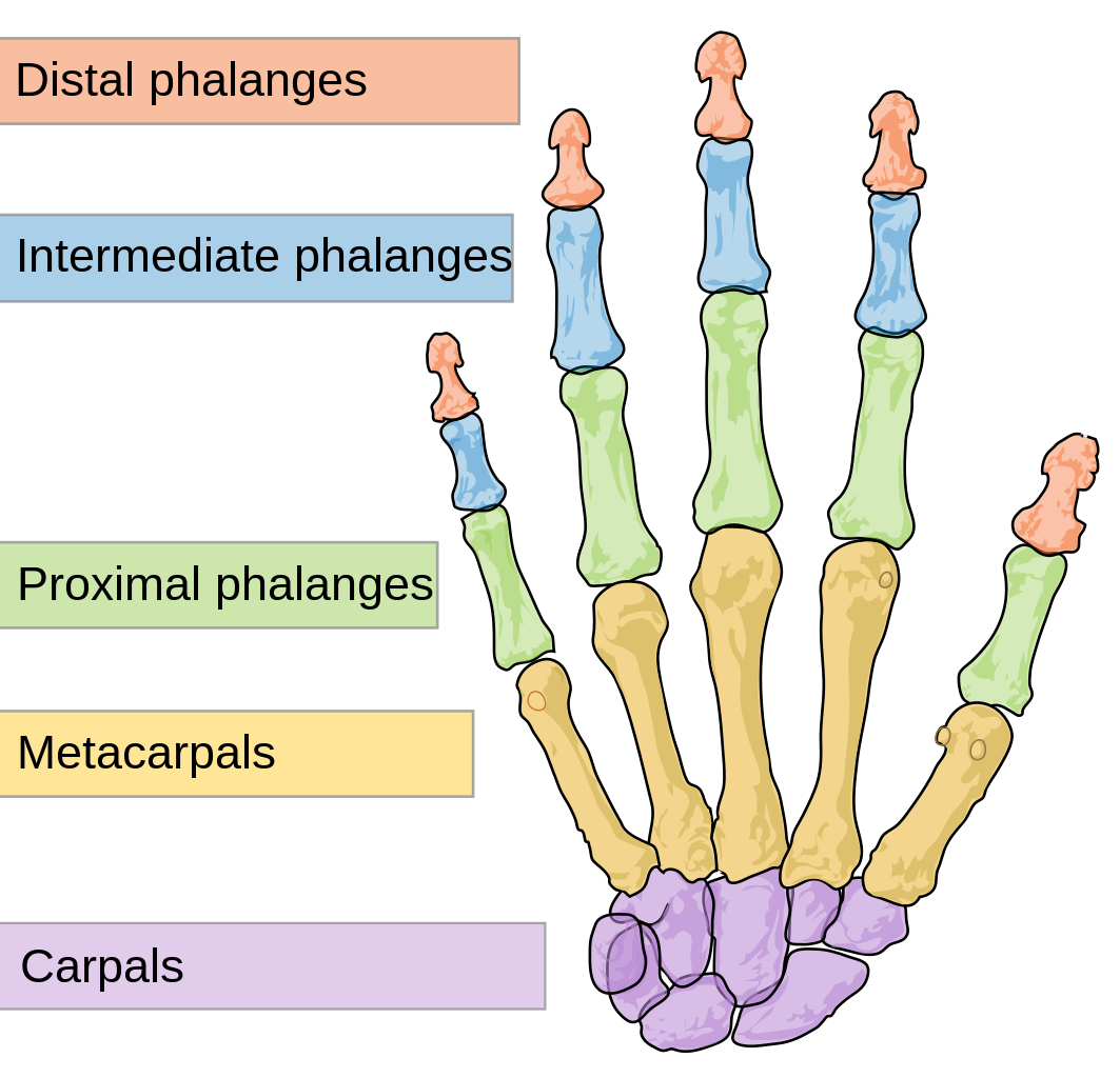
Non-displaced fractures do not require surgery. However, they do produce pain, swelling, bruising, difficultly with movement and grip and also sometimes deformity. Zuccarello's fracture was diagnosed with an x-ray. A regular x-ray is shown below which includes the bone anatomy:


As stated in my previous article, there are four basic types of [hand] fractures:
  • Displaced: the bone breaks into two or more pieces and becomes misaligned. There is a separation between the two fracture faces or the faces are no longer lined up. 
  • Nondisplaced: the bone cracks or breaks but doesn't move and maintains its proper alignment and position (The two fracture surfaces have not moved relative to each other).
  • Comminuted: the bone is broken in several pieces in different places.
  • Open fracture: the bone breaks through the skin (closed is when the bone breaks but there is no puncture or open wound). 
An x-ray showing a displaced fracture of the 2nd, 3rd, and 4th metacarpals. 

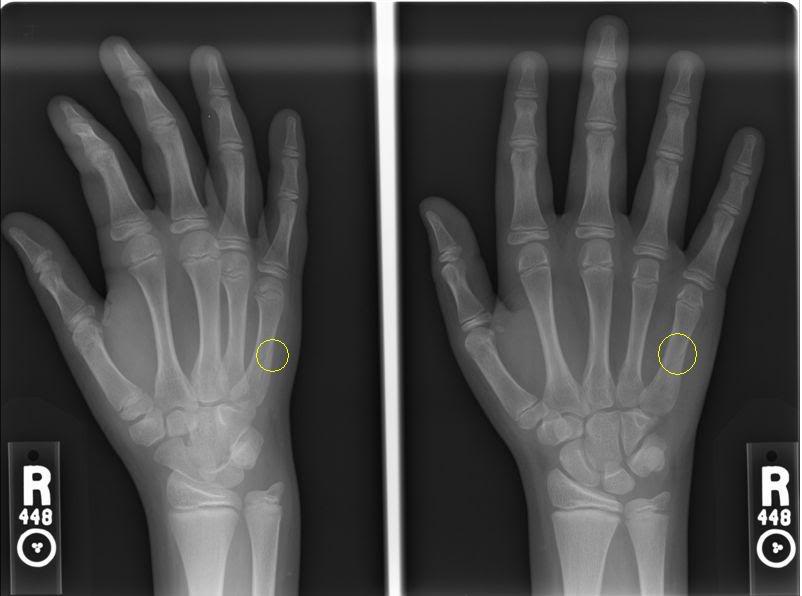
The x-ray image below shows a nondisplaced fracture of the 5th (pinky) metacarpal. The dark line (which is circled), is the fracture. This is the best picture of a nondisplaced fracture I can find. 


The good news for Rangers fans again is that Zuccarello's fracture is nondisplaced and will not require surgery. In cases like this when the bone fragments of the fracture haven't displaced much, or if the break is located in the middle (shaft) of the bone, a specially designed cast (or splint) will be used to hold the bone fragments in place and allow them to heal. Follow-up x-rays will be ordered to ensure that the bone is healing in a good position and hasn't slipped out of alignment. The length of immobilization and healing time depends on several factors (location of the fracture, depth of the fracture, possible injury to the surrounding soft/connective tissue and/or ligaments).

Unfortunately, the quick, less than two-week return for Zuccarello honestly seems unlikely. Hand fractures usually take 3-4 weeks to heal at the very least. On top of that, Zuccarello has broken his left hand previously which is not good news. His previous fracture was fairly serious, and finished his season, sidelining him for 4-6 weeks and not letting him participate in the 2011 World Championships for Norway.

Nondisplaced fractures are a common injury and not too complicated. Zuccarello will fly home and probably see a hand specialist after seeing the Rangers doctor. A cast will be placed (if it already hasn't been), and Zuccarello will rest until fully healed.

Monday, February 17, 2014

Henrik Zetterberg - Spinal Disc Herniation

THE PLAYER 

Henrik Zetterberg, captain of the Detroit Red Wings, was at the Sochi Winter Olympics as captain of Team Sweden. Unfortunately, his tournament ended after just one game due to "devastating back pain." Zetterberg, who has struggled with back injuries all season (has missed 13 games), scored the winning goal against the Czech Republic on Wednesday but woke up on Thursday with a pain in his back that he described as "20 times worse than anything he has previously felt." Zetterberg withdrew from the tournament and is hoping to fly home to Detroit on Tuesday to see a doctor. Fellow Red Wing Niklas Kronwall took over as captain of Team Sweden.


The official word is that Zetterberg has a herniated disc in his back and won't be ready for when NHL games start again after the Olympic break ends. Zetterberg has been unable to travel or even get out of bed due to the pain in his back. 

ANATOMY OF THE SPINE

The vertebral column, also known as the backbone or spine, is a column consisting of 24 articulating vertebrae as well as nine fused vertebrae in the sacrum (five) and the coccyx (four coccygeal bones) which form the tailbone or back wall of the pelvis. The column, which is situated in the dorsal (fancy anatomy term for back) aspect of the human torso, houses and protects the spinal cord in its spinal canal. 
  

Three regions of the vertebral column comprise the 24 articulating vertebrae and they are grouped under the following names:
  • Cervical: 7 vertebrae which are designated/numbered C1 through C7 from top to bottom. The cervical vertebrae are small and delicate. While there are seven cervical bones, there are eight cervical spinal nerves. C1 is called the atlas and C2 is called the axis, these top two vertebrae allow the neck and head so much movement. 
  • Thoracic: the next 12 vertebrae which are designated T1 through T12. The surfaces of the thoracic bones articulate with the ribs, this connection not allowing for much movement. 
  • Lumbar: containing the last five individual vertebrae designated L1 through L5. As the vertebrae of the lower back, they are robust because they support much more weight than the other vertebrae. The lumbar bones also allow significant movement. 

There are 30 spinal cord nerve segments. The nerves that make up the spinal cord exit the vertebrae through the intervertebral foramina and proceed on to different parts of the body that they are responsible for. 
  • C1-C6: Neck flexors
  • C1-T1: Neck extensors
  • C3, C4, C5: Supply diaphragm
  • C5,C6: Shoulder movement, raising arm, flexion of elbow, C6 externally rotates arm
  • C6,C7: Extends elbow and wrist, pronates wrist
  • C7, T1: Flexes wrist, supply small muscles of the hand
  • T1-T6: Intercoastals and trunk above the waist
  • T7-L1: Abdominal muscles
  • L1, L2, L3, L4: Thigh flexion
  • L2, L3, L4: Thigh adduction, extension of leg at the knee.
  • L4, L5, S1: Thigh abduction, extension of toes
  • L5, S1, S2: extension of leg at the hip, plantar flexion of foot, flexion of toes
  • L4, L5, S2: Flexion of lag at the knee (hamstrings)
  • L4, L5, S1: Dorsiflexion of foot


The vertebrae are separated and cushioned by discs. Each cartilaginous disc forms a joint which allow slight movement of the vertebrae, allowing you to bend and reach. Additionally they put space between your vertebrae but act as a ligament to hold them together. These discs consist of an outer fibrocartilage layer and a jelly-like center which act as a shock absorber. 


THE INJURY - WHAT IS A HERNIATED DISC?

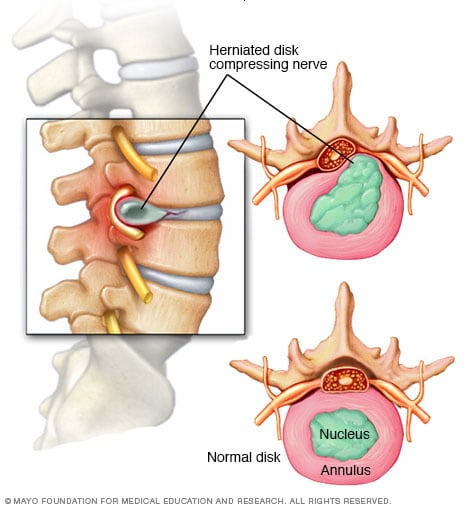
Sometimes referred to as a slipped disc, a spinal disc herniation is when a tear in the outer, fibrocartilage ring allows the soft, jelly-like central portion of bulge out beyond the damaged outer ring. A herniation of the disc is usually a further development of a disc protrusion, a condition in which the outer layer/ring is still intact but bulges when the disc is under pressure. A protrusion is less severe because unlike a herniation, none of the jelly-like nucleus of the disc escapes out of the annulus. 

Disc herniation may be caused by trauma or a single excessive strain. However, they are usually attributed to general wear an tear and age-related degeneration of the outer cartilage ring. As you age, the discs begin to dry out and weaken, shrinking the discs and causing the spaces between the vertebrae to get narrower. This is disc degeneration and is natural, also because ligaments holding the disc in place weaken, a relatively minor strain/twisting motion can cause a disc to rupture. Research has also shown that certain individuals may be vulnerable to back problems and have a predisposition for herniated discs. For example, mutation in genes coding for proteins involved in the regulation of the extracellular matrix, such as MMP2 and THBS2, have been demonstrated to contribute to lumbar disc herniation. Additionally, disc herniation and the associated neurological syndrome has been lined to a human chromosomal region, found on chromosome 6q23.3-q24.1. 

Symptoms 
Symptoms vary greatly depending on where the herniation has occurred and its size. The most common initial symptom is lower back pain. However, if the herniated dic is pressing on a nerve, there may be immense pain, numbness, or weakness in the area of the body to which the nerves of that location innervate. In some patients, a tear of the outer ring and a herniation may cause an immense pain due to chemical inflammation. If there is a herniation in the cervical spine, neck pain can occur which can also radiate throughout the whole upper body. 

One of the most common symptoms of a herniated disc is sciatica. This occurs when there is a herniated disc in the lumbar portion of the spine which causes pressure on one of several nerves that contribute to the sciatic nerve. This can cause pain, weakness, tingling, and numbness that radiates from the buttocks into and down the leg towards the foot. Sciatica is usually only on one side. 
Diagnosis 
To diagnose a disc herniation, the doctor will start with a routine physical examination and will include a straight leg raise (SLR) test. During the SLR test, the doctor will lie you on your back and lift your affected leg (if you're experiencing leg weakness/pain due to sciatica). If you feel pain down your leg when it's being raised, you test positive for a herniated disc. Your doctor will also perform a neurological examination to detect [muscle] weakness or sensory loss. Other diagnostic tests will be performed, specifically imaging to view the spine and the herniation:
  • X-ray: using small doses of radiation to produce images of the body, an x-ray will be obtained to search and rule out other potential causes of pain such as tumors, infections and fractures.
  • CT Scan: A diagnostic image created using a combination of computer technology and x-rays, a CT scan can show the shape and size of the spinal canal, its contents and the surrounding structures. 
  • MRI: uses magnets and computer technology to produce a 3D diagnostic image which can show the spinal cord, nerve roots and surrounding areas. Can depict enlargement, degeneration and show narrowing of the spinal canal caused by a herniation. 
An MRI of the lumbar spine. Normal.

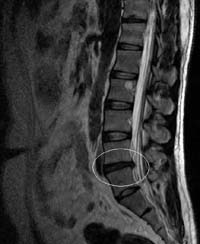
An MRI of the lumbar spine showing a lumbar disc herniation between L4 and L5. 
  • Myelogram: an injection of contrast dye into the spinal canal/surrounding cerebrospinal fluid spaces followed by an x-ray or CT scan. Shows pressure on the spinal cord or nerves due to herniated discs and can help pinpoint the size and location of the herniation.
The contrast dye makes the spinal canal clearly visible. 
The arrow points to a herniated disc compressing the spinal nerves. 
  • Electromyogram (EMG): by placing small needles into various muscles and measuring electrical activity, this test measures the electrical impulse (muscle's response) along nerve roots, peripheral nerves and muscle tissue. This indicates ongoing nerve damage and nerve compression.
Treatment
Fortunately, the majority of herniated discs do not require surgery as the herniated disc will slowly improve over a period of several days to a few weeks, with most patients becoming symptom free in 3-4 months. A doctor will advise bed rest (few days) to calm severe back pain and then advise a low, painless activity level which will help the spinal nerve inflammation to decrease. doctors will also treat a herniated disc with nonsteroidal anti-inflammatory drugs (NSAID) which will relieve mild to moderate pain (medicines like ibuprofen and naproxen). In some cases, doctors will perform an epidural steroid injection. During this procedure, a spinal needle will inject steroid medication into the back under guidance of an x-ray. The directed medication will be applied at the site of the herniation and will reduce local inflammation. These injections have been proven to be successful and reduce symptoms in 42-56% of patients who have not improved in >6 weeks of nonsurgical care. Physical therapy will also be advised along with specific exercises to strengthen the patient's lower back and abdominal muscles. This therapy can also include pelvic traction, massages, ice/heat therapy, ultrasound, electrical muscle stimulation and stretching exercises. 

In the cases of incapacitating back pain and when conservative nonsurgical treatment options do not work, doctors will recommend surgical treatment. Sometimes, surgery is recommended if the herniation is severe and pressing on the spinal cord. However, as always, surgery is a big decision and options must be weighed. 

The most common surgical procedure for a herniated disc in the lower back is a lumbar microdiscectomy. This surgery involves removing the herniated part of the disc along with any fragments which are pressuring the spinal nerve. To perform a discectomy, the surgeon must first put the patient under general anesthesia and perform a laminectomy. A laminectomy is the surgical removal of most of the bony arched lamina of a vertebra. The lamina is removed to allow access to the disc and the nerves, and the disectomy proceeds either as an open procedure, percutaneously (through a small incision), or as a microdisectomy as stated before which uses special microscopes to allow the surgeon to make precise incisions and cause less damage to the surrounding tissues. After the incision is made, the muscles are moved to the side so the surgeon can see the vertebrae and using a retractor, the nerve roots are held to the side as well. The surgeon then uses a rongeur to remove the disc tissue pressuring the spinal nerve. If a disc is removed, the spine must be stabilized, usually through spinal fusion (bone is grafted onto the spine, creating a solid union between two or more vertebrae) or if you're a special candidate, through artificial disc insertion (CHARITE or PRODISC-L). 

The hospital stay following surgery is usually one to two days and the following recovery can take six weeks or more. Post-surgery, patients must undergo physical therapy to increase strength in the lower spine. An eight-year follow-up study published in Spine showed that patients with herniated discs in the lumbar spine that had surgery, had greater long-term improvement in pain, functioning, and disability when compared to nonsurgical treatment. For example, on a 100-point pain scale, pain scores averaged about 11 points lower in the group that obtained the surgery when compared to the non-surgical group. This occurred in measures of physical functioning as well. However, the study also showed that nonsurgical treatment can provide lasting benefits.

Update: u/big_phat_gator (heh) on r/detroitredwings asked a good question regarding the risks of surgery. I thought it was a good question and warranted the answer being posted here:

Discectomy is a relatively routine and safe surgery. However, every surgery has general associated risks such as:

  • Adverse events due to Anesthesia (very small risks of breathing problems, other issues)
  • Bleeding
  • Risk of infection (small since doctors now preemptively prescribe and provide antibiotics post-surgery)
Specific risks related to a discectomy procedure: 
  • Nerve damage
  • Dural leak - loss of cerebrospinal fluid (CSF - surrounds the nerve in the spinal canal) if the thin nerve root canal is accidentally ruptured/opened. Usually causes headaches but is resolved in a matter of days
  • Nerve compression causes by a hematoma - blood collecting around the nerves post-surgery can pressure the nerve causing pain/weakness
  • Relapse, or another disc herniation occurring after surgery.  
Zetterberg will fly home to Detroit and see a doctor. Since he's suffered from this before and underwent nonsurgical treatment, surgery may be necessary but its unclear at this time. Lumbar disectomy is one of the most common procedures in the United States and after the 6-week recovery period and therapy, Zetterberg should be much better and perhaps return for the playoffs (if the Red Wings are in contention). However, his back problems have been nagging which is not good and his age may complicate things. Look out in the coming week for more news on Zetterberg and his decision. 


Thursday, February 13, 2014

Frans Nielsen's Fractured Hand

THE PLAYER AND INJURY

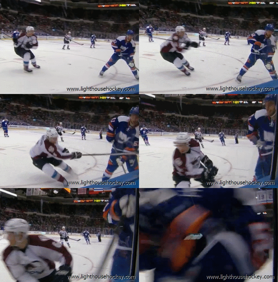
Last Saturday on the last game before the Olympic break, Frans Nielsen of the New York Islanders suffered a broken left hand. The break was directly caused due to a two-handed slash delivered by Colorado Avalanche defenseman Erik Johnson which earned him a two-game suspension. The suspension video is here.

Johnson delivered a reckless, vicious slash that caught Nielsen across the thumb. Nielsen did not return to the game. Gif below courtesy of Lighthouse Hockey.


Frans may be a Danish God, but that hurts. 

The word from Islanders beat writer Arthur Staple is that Nielsen suffered nondisplaced fracture in his left hand and may be out a month.

NIELSEN'S FRACTURE, RECOVERY, AND PROGNOSIS

Fractures in the bones of the hand are actually a fairly common injury in winter sports. For example, skier's thumb, a fracture of the thumb leading to damage of the ulnar collateral ligament (UCL) is the most frequent upper extremity injury second only to knee injuries in downhill skiing. The hands are vulnerable in sports such as hockey to hooks, slashes and falls/crashes to the ice or boards. There are several bones in the hand vulnerable to fracture; the phalanges of the five digit fingers, the long mid-hand metacarpal bones, or the carpal bones of the wrist/lower hand (eg scaphoid, lunate, capitate, etc.).


A fracture is a break in the bone and presents symptoms including pain, swelling, bruising, difficulty with movement, and deformity (depending on the nature of the fracture). To diagnose a hand fracture, usually only a  plain x-ray. A CT scan, MRI, or bone scan could also be used if the fracture is complex or difficult to visualize on an x-ray and also if there is a concern for ligament damage.

An x-ray showing a fracture of the 2nd, 3rd, and 4th metacarpals. 

There are four types of [hand] fractures:
  • Displaced: the bone breaks into two or more pieces and becomes misaligned. There is a separation between the two fracture faces or the faces are no longer lined up. 
  • Nondisplaced: the bone cracks or breaks but doesn't move and maintains its proper alignment and position (The two fracture surfaces have not moved relative to each other).
  • Comminuted: the bone is broken in several pieces in different places.
  • Open fracture: the bone breaks through the skin (closed is when the bone breaks but there is no puncture or open wound). 
The x-ray image below shows a nondisplaced fracture of the 5th (pinky) metacarpal. The dark line (which is circled), is the fracture. 


The good news for Islanders fans is that Frans Nielsen's fracture is nondisplaced and will not require surgery. In cases like this when the bone fragments of the fracture haven't displaced much, or if the break is located in the middle (shaft) of the bone, a specially designed cast (or splint) will be used to hold the bone fragments in place and allow them to heal. Follow-up x-rays will be ordered to ensure that the bone is healing in a good position and hasn't slipped out of alignment.

If a fracture is displaced and unstable, surgery may be necessary to set the bones back into alignment and stabilize them with screws, pins, or plates, ensuring proper healing. Surgery of the hand may involve a region, or general anesthetic depending on the location and severity of the injury. If a fracture can be aligned and stabilized without an incision, it's called a closed reduction. However, if a fracture requires an incision for manipulation and stabilization of the fractured bone (with wires, screws, plates, etc) it is known as an open reduction, internal fixation (ORIF). In the event of a comminuted fracture where the fragments are shattered or bone becomes missing, a bone graft may be necessary. Post-operative management requires a splint/cast immobilization.

The length of immobilization and healing time depends on several factors included but not limited to: location of the fracture, depth of the fracture, injury to the surrounding soft/connective tissue, and injury to ligaments. Possible and common complications following a fracture include stiffness, soreness, slow-healing and mis-aligned healing.

The four-week/month time frame stated by Arthur Staple seems fairly standard. He's expected to miss the first week of Islanders games following the Olympic break. The only issue seems to be the location of his injury (possibly the thumb), an important and complex area of the hand. However, it seems like it'll be an easy recovery for Nielsen who will miss minimal time thanks to the two-week break. Let's hope for a full recovery, because Nielsen needs his hands healthy to pull off his patented Danish Backhand of Justice (DBOJ) as shown below.


Saturday, February 8, 2014

Kris Letang's Stroke

THE PLAYER AND TIMELINE

Kris Letang hasn't played since January 27th at Buffalo. The 26 year-old and Norris Trophy Finalist from the 2013 season signed an 8-year, $58-million year this past summer and has battled with injuries all season (including an infection in his elbow and subsequent operation). On January 30th, Letang was scratched and Penguins coach Dan Blysma was quoted saying Letang was ill. Letang missed the Penguins next game against the Coyotes on the 1st of February due to "being sick" and on the 3rd, the Sporting News published that Letang would miss his third consecutive game and will be evaluated further.
"Asked whether it's a long-term or serious issue, Blysma said it was "uncertain."
Three days later on the 6th, the Penguins announced on Twitter:

Later that evening, Rob Rossi who covers the Penguins for the Pittsburgh Tribune-Review, dropped a bomb on twitter and via the Tribune website saying:
"Kris Letang's mystery illness is not believed life threatening, but the Penguins are not sure when he will play again this season...There were concerns last week that Letang's season - if not his career - could be in jeopardy because of the illness, sources said."
 This morning, Letang's mystery illness was revealed, and it was shocking.
Kris Letang and the Penguins organization have been very open about his condition on twitter and their site, with the hope of sending awareness to others. They deserve recognition for this (especially considering how taboo NHL organizations are about injuries) and everyone in the hockey community wishes Letang a healthy recovery.

LETANG'S STROKE, CAUSES, EVALUATION AND PROGNOSIS

While in Phoenix with his team, Letang experienced dizziness and nausea on the morning of the 29th of January. Letang was kept out of practice and the Penguins games by team doctors and underwent testing. He was diagnosed with having a stroke on the 1st which was confirmed on the 6th after undergoing an array of tests to determine how damaging the stroke was and its origin.

Diagnosis
Team doctors reviewed Letang's history and gave him a physical exam. During the physical exam, the physician will check the player's mental alertness, coordination, and balance. Along with checking for those neurological symptoms, the physician will check for numbness or weakness in the player's face and extremities. To cover all the bases, the physician will also look for signs of carotid artery disease which is a common cause of ischemic stroke by listening to your carotid arteries with a stethoscope. Listening to the arteries can point to changed or reduced blood flow to the heart due to plaque buildup in the carotid arteries.

After the physical exam, the doctor will order an array of diagnostic tests and procedures to verify the diagnosis of stroke. The various tests which could have been ordered for Letang are listed below:

  • Brain Computed Tomography (Brain CT Scan): A CT scan uses a combination of x-rays and computer technology to take detailed pictures of the patient's brain. The scan can show if there is bleeding in the brain or damage to the brain cells. 
  • Brain Magnetic Resonance Imaging (MRI): An MRI uses magnets and radio waves to create detailed pictures of the brain. MRI's can detect changes in brain tissue and damage to brain cells. An MRI shows the brain tissue in more detail than a CT scan. 
Left: CT scan slice of the brain showing a right-hemispheric ischemic stroke.
Right: MRI showing damaged brain cells due to a left-hemispheric stroke.
  • Arteriogram: Either a CT arteriogram (CTA) or a Magnetic Resonance Arteriogram (MRA) can show the blood vessels in the brain and give doctors information about the site of a blood clot and how the blood is flowing through a patient's brain. 
  • Carotid tests: Doctors can order a Carotid Ultrasound or a Carotid Angiography to get pictures of the carotid arteries. The ultrasound uses sound waves to create pictures while an angiography uses dye and x-rays. The carotid arteries supply oxygen-rich blood to the brain and can be narrowed or blocked by plaque build-up. 
An angiogram showing a major blood vessel which is blocked and caused a stroke.
 Arrow indicates the location of the blockage. 
  • Heart tests: To determine and detect if heart problems may have led to a stroke, a doctor may order an EKG (Electrocardiogram) or an Echo (Echocardiography) test. An EKG will record the heart's electrical activity to determine if the heart has an irregular rhythm such as atrial fibrillation. An echo uses sound waves to create pictures of the heart. This will be discussed later because it's particularly important in Letang's case. 
  • Blood tests including blood glucose tests (low blood glucose levels may cause similar symptoms), platelet counts (abnormal platelet levels may be a sign of a bleeding disorder), PT/PTT tests to determine blood clotting and toxicology screen to determine if drug use was a cause.
Diagnosing a stroke can be difficult and since it's so serious, it's important to diagnose correctly. A recent study at the Wayne State University-Detroit Medical Center Stroke Program found that 1/7 young stroke victims were given a misdiagnosis of vertigo, migraine, drug/alcohol abuse, seizure, or other problems and sent home without proper treatment. This is partially due to the lower risk for stroke among young people; people aged 65+ have a risk of 30-50/1,000 while people 45 and younger have a low risk of 1/1,000. 

There are two types of strokes - ischemic and hemorrhagic. An ischemic stroke is caused by an interruption in blood supply to a certain part of the brain, usually from a clot. This is the type of stroke Letang suffered, but thankfully he just had mild symptoms that were diagnosed days later. A hemorrhagic stroke is caused by bleeding into the brain. Additionally, it's important to also determine if a stroke occurred or a transient ischemic attack (TIA). A TIA is different than a stroke, unlike a stroke, a TIA does not cause brain tissue to die. A TIA is caused by a brief pause in blood flow to part of a brain due to a temporary or partial blockage. After a TIA, the blockage breaks up quickly and dissolves but serves as a warning sign as more than 10% of people who have a TIA will have a stroke within 3 months with half of these strokes happening 48 hours or fewer after a TIA (NIH)

In Kris Letang's case, it was revealed that he suffered a stroke most likely due to a congenital (meaning present from birth) heart defect (CHD), specifically a small hole in his heart. Heart defects are the most common birth defects with approximately 9 in 1,000 people being born with a CHD. Many defects don't need treatment, but if they create complications they may require medication or surgery. The heart has two sides, separated by an inner wall called the septum. The left side of the heart receives oxygen-rich blood from the lungs and pumps it to the body while the right side receives oxygen-poor blood from the body, pumping it to the lungs. The septum prevents the mixing of blood. A hole in the septum between the heart's two upper chambers is called an atrial septal defect (ASD) while a hole in the septum between the heart's two lower chambers is called a ventricular septal defect (VSD). ASDs and VSDs allow blood to pass from the left side of the heart to the right side, mixing the oxygen-rich and oxygen-poor blood which can result in oxygen-rich blood being pumped to the lungs instead of the body in need of the oxygen.  


After his symptoms and cryptogenic stroke diagnosis (meaning a stroke of unknown cause), doctors most likely ordered an echo for Letang to rule out or determine a CHD. Transoesophageal echocardiography (TOE) is regarded as the imaging procedure of choice to diagnose a CHD, specifically a patent foramen ovale, or PFO. Pictured below is an image of a TOE from a patient with a PFO. Source: Pinto. 

TOE of a patient with a PFO, shows a wide separation in the inter-atrial septum (arrowed).
Ao, (aortic root); IAS, inter-atrial septum; LA, left atrium; RA, right atrium. 

In approximately 25% of adults, the foramen ovale does not entirely seal (Kumar) which results in a patent foramen ovale (PFO), a type of atrial septal defect. A PFO allows blood that hasn't been cleansed by passing through the lungs to flow through the flap, carrying debris and blood clots that can travel up to the brain, causing a stroke. 

Treatment and prognosis
Patients who have had a stroke and are diagnosed with a PFO are prescribed blood thinners such as aspirin or warfarin (branded as Coumadin) to decrease the chance of another stroke. The drugs don't seal the flap, obviously, but reduce the risk of blood clots which could travel to the brain and cause another stroke. Blood thinners (anti-platelets and anti-coagulants) must be taken daily. As I described in my article on Tomas Vokoun's blood clots, players can not undergo any physical/sport activity while on anti-coagulant medication. Blood thinners increase the risk for bleeding as your blood isn't able to clot at a wound. If a player is accidentally cut or bruised badly, there is a chance of death. 

Open-heart surgery is an option to close the PFO however it is rarely used for people who don't respond to the blood thinner drugs. As it's a major surgery, patients and doctors need to weigh the risks of the surgery with the benefits. During open-heart surgery, the cardia surgeon makes an incision in the chest to reach the ASD and repairs the defect with a specially-made patch to cover the hole. A heart-lung bypass machine is used during the surgery which takes over the heart's pumping action and moves blood away from the heart. While the outlook for patients who have open-heart surgery is pretty good and complications are rare, open-heart surgery is a huge procedure. One of the more common complications is pericarditis, or inflammation of the outer lining of the heart which causes fluid to collect around the heart which can be treated with medication. 

Another option of treatment is a minimally-invasive, catheter-based procedure to close the PFO. This percutaneous PFO closure is favored over open-heart surgery due to being a safer and less-invasive/painful procedure. Major periprocedure complications (i.e. death, hemorraging, need for further surgery, etc.) occurs in about 1.5% of patients with minor complications (bleeding, complications with the device, etc.) occurs in about 7.9% of cases in a retrospective study of patients that received a PFO closure (Landzberg et al). 

In a catheter-based procedure, a patent forament ovale closure device is guided to the PFO via a catheter. The device becomes a permanent implant that will close the PFO in the heart wall as heart tissue grows around the implant, sealing the hole. The catheter is inserted into a large vein using a small incision in the inner thigh and is advanced into the heart. The closure device is moved through the catheter to the heart to the location of the PFO and once in the correct location, the device is placed so that it straddles each side of the hole and the catheter is removed. The cardiac catheterization procedure for a PFO closure typically takes 1 to 2 hours and requires 6 hours of bed rest post-surgery, meaning the surgery is sometimes an outpatient procedure, allowing patients to leave the hospital the same day. After surgery, tests will be done to ensure proper placement of the implanted closure device. Patients usually can't undergo physical activity for up to a week and continue to take blood-thinning medications along with antibiotics to prevent infection. 

The devices are amazing bits of technology and resemble tiny umbrellas. One device called the HELEX Septal Occluder consists of a circular wire frame made of a nickel-titanium metal alloy covered with a thin membrane layer of Gore-Tex (which has been used in open-heart surgery for > 20 years). Once the device is passed through the catheter, it opens up to form a circular disk that covers the hole as the device is slightly larger than the hole. Another (slightly bulkier) device is the Amplatzer Septal Occluder which consists of two expandable discs made of nickel-titanium metal alloy. The discs have a polyster mesh incorporated inside to enhance elimination of flow across the hole. 


A HELEX Septal Occluder device for PFO Closure. 

As of right now, Kris Letang is on blood-thinning medication (aspirin most likely), and is out for a minimum of six weeks. It's uncertain whether or not he will pursue surgery. His stroke is a scary thought, and everyone wishes him the best. As more information comes out, we'll see how Letang progresses. As stated on the Penguins site, doctors don't believe it will end Letang's career and Letang hopes to play again. 


SOURCES: 

F J Pinto. When and how to diagnose patent foramen ovale. Heart. 2005 April; 91(4): 438-440. 

Khairy P, O'Donnell CP, Landzberg MJ. Transcatheter closure versus medical therapy of patient foramen ovale and presumed paradoxical thromboemboli: a systematic review. Ann Intern Med 2003;139:753-760. 

Kumar, Vinay (2007). Robbins Basic Pathology (8th ed. ed.). Philadelphia: Saunders/Elsevier. p. 384. 

Monday, February 3, 2014

Reviewing Concussions: Lubomir Visnovsky returns after his post-concussive troubles. What is prolotherapy?

Lubomir Visnovsky, 37, returned January 27th in a 6-3 Islanders loss against the Boston Bruins. The veteran defenseman was absent from the lineup for three months, missing 46 games, more than half a season.

Visnovsky was injured on October 19th in the first period of a game against the Carolina Hurricanes on a seemingly innocent hit by forward Riley Nash. Contrary to original belief, many believed Visnovsky was injured on a check by Hurricanes forward Radek Dvorak, which is shown below in gif format courtesy of of Islanders Point Blank. After the Dvorak hit, Visnovsky made it to the bench on his own but left the ice hunched over during the next TV timeout (Islanders benches at Nassau Coliseum don't have direct access to the locker room) and didn't return. Visnovsky wanted to make clear that although he left after the Dvorak hit, it was the hit earlier in the game by Nash.


In an article in the New York Post, Visnovsky described to writer Brett Cygralis that he immediately knew he had a concussion because of his previous concussion history. Visnovsky's last concussion was during the last game of the regular season in 2012 when he was a member of the Anaheim Ducks. Additionally, Visnovsky suffered 4 previous concussions while a member of the Los Angeles Kings and his hometown Slovakian team, Bratislava Slovan.

However, according to Visnovsky, this concussion lingered and kept him out of commission much longer than most. Is it the cumulative effect of his previous concussion history? Does it have to do with his age? Possibly, they all can be considered factors, but the bottom line is we don't know. Additionally, a players post-concussion management and symptoms can be affected by their social situation. Does Visnovsky have a strong social structure at home on Long Island? There are several factors.

In the NY Post article, it's reported that Visnovsky visited renowned concussion specialist, Dr. Jeffrey Scott Kutcher. Kutcher is very well known in the world of neurology, an has worked with NHL players such as Mark Staal, the NHLPA, and will be traveling to Sochi as Team USA's neurologist/medical consultant. Kutcher is well respected in the industry and since he sees such high-profile patients (mostly, if not all athletes) and participates in numerous concussion research studies, he's able to provide new and unique care for patients struggling with concussions.

Visnovsky's concussion lingered and he was unable to return to play or follow a specific timeline to recovery. Visnovsky would "wake up with a migraine," and it would last all day, frustrating him. However after seeing Dr. Kutcher on New Year's eve, Visnovsky felt much better and was able to return in less than a month. So what therapy did he undergo?


PROLOTHERAPY- WHAT IS IT ACTUALLY? 

It's reported that Kutcher gave Visnovsky two injections to the back of his neck as part of a treatment known as prolotherapy. As Cygralis says in the NY Post article, prolotherapy is meant to avoid surgery and help the nerves of the neck "communicate better with the brain, lessen pain (including headaches) and increase mobility."

When a player sustains a concussion, or any form of traumatic head injury, multiple things happen during the mechanism of injury. Not only is there significant trauma to the skull which damages the brain, a significant whiplash injury can be sustained to the neck and base of the skull. The skull base is an important area, containing the brainstem, the posterior part of the brain which is structurally continuous with the spinal cord. The brainstem is extremely important, playing an important role in the regulation of cardiac and respiratory function (why you don't have to think about breathing or heart beats) and providing the passage for nerve connections of the motor and sensory systems from the spinal cord to the cerebrum (brain). Included in these systems are tracts such as the corticospinal tract or motor system (controlling things such as balance and coordination) and the spinothalamic tract (pain, temperature, itch, etc.). In addition, the brainstem also regulates the central nervous system (CNS) and is pivotal in regulating the sleep cycle (many concussion patients struggle with sleep).

Injury to the skull base and upper neck area may change the relationship between the brainstem, cerebrum, and cervical canal, causing increased pressure on nerve control. Those that use prolotherapy on their patients, believe that this increased pressure leads to many of the symptoms of post-concussion patients.

Prolotherapy is a method of injection treatment designed to stimulate healing which is designed to be used for musculoskeletal pain that has occurred for loner than 8 weeks (Hackett et al.). Essentially, prolotherapy works by raising the growth factor levels or effectiveness to promote tissue repair or growth and is considered a long-term solution (Reeves). The treatment is based on the premise that chronic musculoskeletal pain is due to inadequate repair of fibrous connective tissue, resulting in ligament and tendon weakness or relaxation, also known as connective tissue insufficiency (Hackett et al.).

Since ligament and tendon tissues have poor blood supply, they take longer to heal that other tissues. Additionally, it's been estimated that the usual best result of a completed connective tissue repair process is only 50-60% of pre-injury tensile strength (Andriacchi et al.). Over time, multiple injuries (additional trauma) and interfering factors such as stress and lack of sleep may interfere with the healing process.

Prolotherapy is designed to cause a temporary, low grade inflammation at the site of ligament or tendon weakness, which is supposed to "trick" the body into initiating a new healing process in the affected area. Inflammation activates fibroblasts to the area, which are a type of cell that synthesizes collagen, thereby reinforcing connective tissue (Reeves).

Prolotherapy has been used in the United States for musculoskeletal pain since the 1930s, but has always been considered an alternative therapy despite as many as 450,000 Americans having undergone prolotherapy.

In fact, Visnovsky isn't the first NHL player to receive prolotherapy. Simon Gagne who suffered multiple concussions late in his NHL career, underwent prolotherpy with Dr. Greenberg in New Jersey. In the video below, both Dr. Greenberg and Gagne discuss the process.


CONCLUSION/TLDR

Visnovsky was concussed and after several months of no significant improvement and a difficult return to play management, he opted for an alternative therapy known as prolotherapy. Now is prolotherapy legit? Honestly, many doctors haven't even heard of it. Concussion is a difficult injury, accompanied by symptoms associated with unknown causes. Many think somatization is a difficult issue when dealing with post-concussion syndrome. Is it possible prolotherapy helps? Yes. Is it possible it's also a placebo where the patient thinks they're receiving treatment and actually recover on their own? Possibly. 

The bottom line is Visnovsky is back, which is good for the Islanders.  


SOURCES:

Andriacchi T, Sabiston P, DeHaven K, et al. Ligament: Injury and Repair. Acta Rheum Scand. 1956. 2:109-116. 

Hackett GS, Hemwall GA, and Montgomery GA. Ligament and Tendon Relaxation Treated by Prolotherapy. (1956 First Edition; Charles C. Thomas, Publisher). Fifth Edition. Gustav A. Hemwall, Publisher. Institute in Basic Life Principles. Oak Brook, IL. 1991. 

Reeves KD. Prolotherapy: Basic Science, Clinical Studies, and Technique. Lennard TA (Ed) Pain Procedures in Clinical Practice, 2nd Ed. Hanley and Belfus. Philadelphia. 200. pp 172-190.